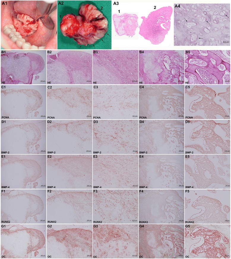
Fig. 2.

BPOP. A1 Sublingual ulceration with a whitish calcified mass. A2 Removed mass showing partial bluish color (arrows). A3 BPOP specimen was composed of cartilaginous (1) and osseous (2) tissue. A4 Bizarre chondrocytes (arrows) in cartilaginous tissue. B HE stain. B1–B3 (area 1 of A3). B4, B5 (area 2 of A3). B1 and B3 show core cartilage covered with thick perichondral fibrous tissue. B4 and B5 show anastomosing trabecular bone centered from cartilaginous tissue, mimicking endochondral ossification. C–G IHC stains with no background stain. C PCNA. D BMP-2. E BMP-4. F RUNX2. G OC
