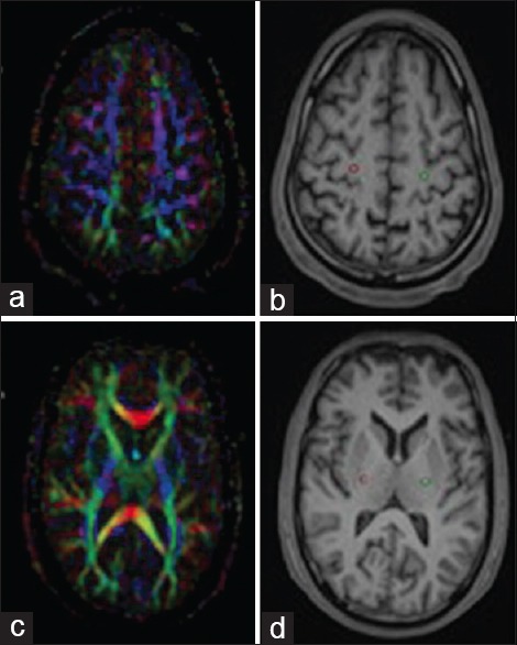
Figure 1.

Placement of 3 mm3 sized regions of interest (ROI) in primary motor cortex (a) and mid-portion of the posterior limb of the internal capsule (c). ROIs were placed on color coded fractional anisotropy maps which were automatically transferred to fused T1 MPRAGE anatomical images (b and d)
