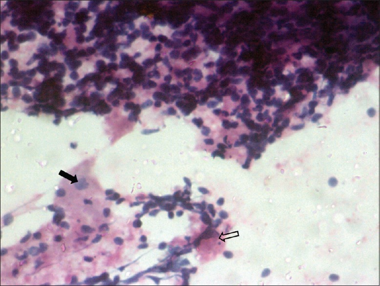
Figure 3.

Papanicolaou staining ×20 of cervical lymph node fine-needle aspiration cytology showing lymphocytes, macrophages, foci of necrosis (shaded arrow), and macrophage engulfing karyorrhectic debris (open arrow) which is characteristic of Kikuchi-Fujimoto disease
