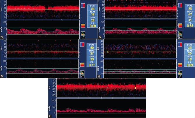
Figure 2.
TCD findings on insonation of Left MCA and PCA. (a) Left MCA (baseline) (MFV: 38). (b) Left MCA (end of breath holding) (MFV: 42). (c) Left PCA (baseline) (MFV: 27). (d) Left PCA (end of breath holding) (MFV: 25). (e) MES left MCA. TCD: Transcranial Doppler, MCA: Middle cerebral artery, PCA: Posterior cerebral artery, MES: Microembolic signal, MFV: Mean flow velocity

