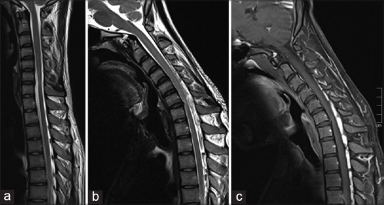
Figure 4.

(a) Sagittal T2-SPACE magnetic resonance imaging (MRI) Cervical spine in neutral position shows of straightening of cervical spine with segmental cord atrophy and intramedullary hyperintensity at C6–C7 level; note the “sand watch” appearance in neutral position with normal cord architecture above and below the atrophy. (b) In flexion position shows multiple large flow voids in the cervico-dorsal posterior epidural space which shows enhancement in postcontrast sagittal T1W MRI (c)
