Figure 1.

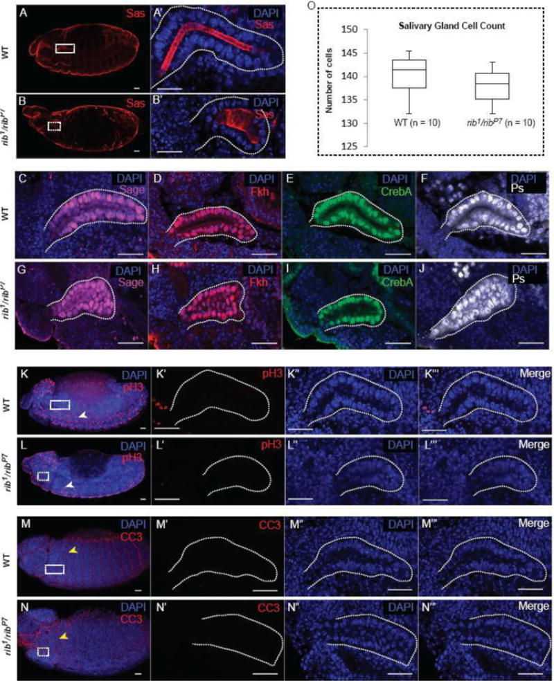
Cell specification, cell division and apoptosis are not affected in rib mutant salivary glands (SGs). (A, B) Wild-type (WT) and rib1/ribP7 embryos stained for the apical cell membrane protein SAS and DAPI show overall embryo morphology. (A′) A high magnification image of the WT SG (white box in A) reveals an elongated SG tube. (B′) A high magnification image of the rib1/ribP7 SG (white box in B) reveals a short, wide SG tube. (C – J) WT and rib mutant SGs were stained with DAPI and four different cell-specification markers: Sage (C, G), Fkh (D, H), CrebA (E, I), and Ps (F, J). (K, L) WT and rib mutant salivary glands were stained with DAPI and phospho-histone H3 (pH3) antiserum to assay for cell division. The white arrowheads indicate one of the many pH3+ neuroblasts undergoing cell division. (K′ – K‴ and L′ – L‴) High-magnification images of WT and rib mutant SGs (white boxes in K and L, respectively) are shown. (M, N) WT and rib mutant SGs were stained with DAPI and cleaved caspase3 antibody to assay for apoptosis. The yellow arrowheads indicate one of the many CC3+ intersegmental surface epithelial cells undergoing apoptosis. (M′ – M‴ and N′ – N‴) High-magnification images of WT and rib mutant SGs (white boxes in M and N, respectively) are shown. (O) Cell count from WT and rib mutants showed no statistically significant difference in the number of cells present in WT versus rib mutant SGs (n = 10 glands). Scale bars: 20 μm.
