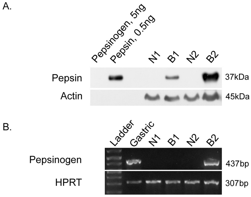
Figure 1. Pepsin protein and transcript were observed in Barrett’s epithelium.
(A) Thirty micrograms (ug) esophageal biopsy lysate was analysed alongside human pepsin 3b and pepsinogen I (positive and negative controls, respectively) via SDS-PAGE/Western blot. Western blot was performed for both pepsin and actin (positive control). (B) RT-PCR was performed on Barrett’s specimens to investigate local pepsin synthesis in Barrett’s tissues. Human gastric cDNA template and hypoxanthine-guanine phosphoribosyltransferase 1 (HPRT) were used as a positive controls. Amplicon corresponding to pepsinogen A (437bp) was detected in human gastric tissue and one of the two Barrett’s esophagus samples. HPRT was detected in all samples (307bp).

