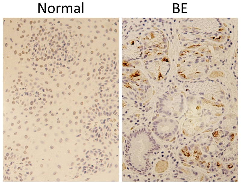
Figure 2. Pepsin immunohistochemistry demonstrates pepsin in Barrett’s epithelium but not in neighboring normal control tissue.
Pepsin immunohistochemistry (brown) was performed on formalin-fixed, paraffin embedded biopsies from BE and neighboring normal tissue from a BE patient. Hematoxylin (purple) was used as counter-stain.

