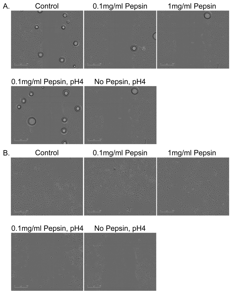
Figure 5. Migration assay 30 minutes and 24 hours following 5 minute acid or acid and pepsin exposure.
Esophageal cells (A) 30 minutes and (B) 24 hours after assay start. Cells were treated with pepsin alone throughout the duration of the assay; acid (pH4) treatment was limited to 5 minutes pretreatment prior to assay start at which time cells were rinsed and normal growth media was replaced. Bubbles observed in (A) are a normal product of media transfer that disappeared by subsequent image collection 2hours post-treatment.

