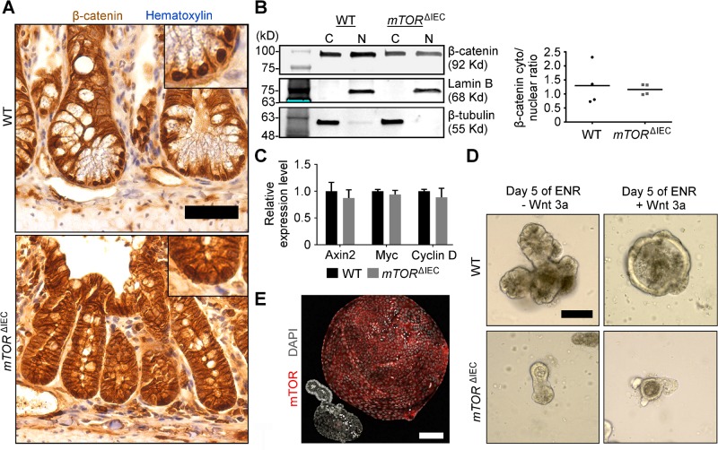
Figure 6.
mTOR is dispensable for small intestinal crypt canonical WNT signaling. A) β-Catenin (brown) IHC staining of WT and mTORΔIEC ileal crypts. Hematoxylin indicates DNA (blue). Insets are magnified equally. B) Representative cytoplasmic/nuclear β-catenin fractionation Western blot of WT and mTORΔIEC ileal crypt cells. β-Tubulin (C, cytoplasmic fraction) and Lamin B (N, nuclear fraction) are shown. Graph shows ratio of cytoplasmic:nuclear β-catenin with lines indicating the mean. P > 0.7. C) Quantitative RT-PCR of WNT signaling targets in isolated WT and mTORΔIEC distal intestinal crypts. Error bars represent sem. D) Representative images of WT and mTORΔIEC enteroids treated with 80 ng/ml WNT3a for 5 d. E) Immunofluorescence (IF) image compares WT vs. mTORΔIEC enteroid morphology after 6 d of 80 ng/ml WNT3a treatment. mTOR is shown in red, and DAPI indicates DNA (gray). A, B) n = 4 mice. C–E) n = 3 mice. Scale bars, 50 μm (A) and 100 μm (D, E).

