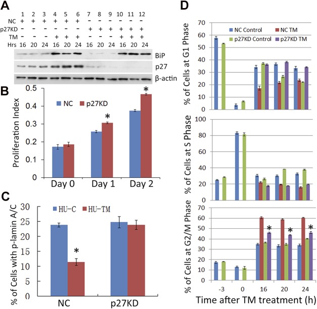
Figure 6.
p27KD abolishes the inhibition of phospho-lamin A/C and G2/M arrest caused by the UPR. A) p27 was knocked down in HLECs using lentivirus-mediated shRNA. The NC and p27KD HLECs were synchronized by HU and treated with TM (Fig. 5A). The levels of p27 and BiP, in the absence or presence of TM, were detected at the indicated time points with Western Blot. B) The growth rates of NC and p27KD cells were determined by the MTS assay, as described in experimental procedures. C) The percentages of phospho-lamin A/C+ cells in the synchronized NC, and p27KD cells were detected by immunostaining 8 h after TM treatment. D) The synchronized NC and p27KD cells were treated with TM or left untreated (Fig. 5A). The cell cycle progressions were determined by FACS analysis. *P < 0.001 vs. TM-treated NC cells at the indicated time points.

