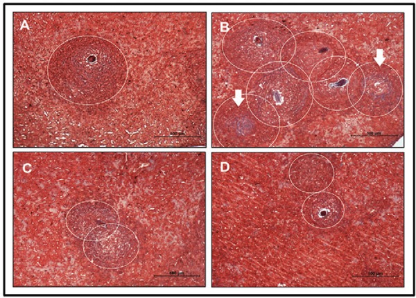
Fig. 2. : histomorphometric study of liver tissue. Analysis of hepatic granulomas in Swiss Webster mice born and suckled from uninfected mothers (control) (A), born from infected mothers (B), suckled by infected mothers (C), and born and suckled by schistosomotic mothers (D) and 69th day post-infection with 80 S. mansonicercariae. The slides were stained with Masson trichrome (selective for collagen) and the analyses were performed on images randomly obtained in 10-20 fields/animal (100X). The histomorphometric study was performed in five animals/group. Circles defining the granuloma size (A, B, C and D) and arrows indicate areas with increased collagen deposition (B).

