
Figure 2.
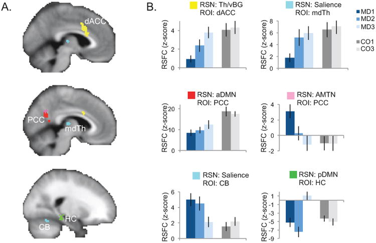
Response to right-unilateral electroconvulsive therapy (ΔECT) within individual resting-state networks of interest (RSNs). A. The results of a linear mixed-effects analysis of ΔECT within each RSN are displayed, restricted to voxels belonging to any RSN (abbreviations given in Figure 1B). Significant clusters are displayed on an average-template brain (pcorr < 0.00625); color indicates the RSN for which the ΔECT effects (described in Figure 1B) were significant (key in B). Effects were significant in dorsal anterior cingulate (dACC), posterior cingulate (PCC), mediodorsal thalamus (mdTh), hippocampus (HC), and cerebellum (CB). B. Mean resting-state functional connectivity (RSFC, z-score) in significant clusters is plotted for each group and time-point at right, where color indicates group and shade indicates time-point (key displayed at right). Error bars mark the standard error of the mean across subjects.