
Fig. 5.
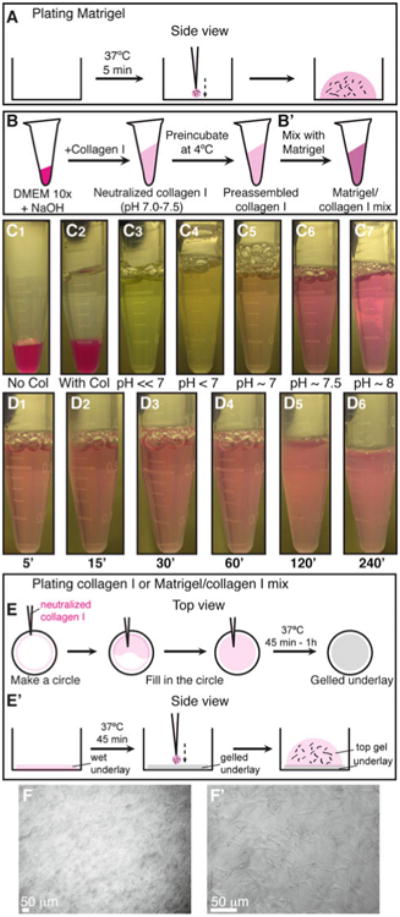
Plating organoids in 3D Matrigel and collagen I. (a) Schematic description of plating organoids in Matrigel. (b–b′) Schematic description of preparing preassembled collagen I (b), which can be used alone or mixed with Matrigel (b′). (c1–c7) Color indicators for the pH of the collagen I solution during neutralization. (d1–d6) Decreasing transparency of the collagen I solution during preincubation on ice. (e–e′) Schematic description of plating organoids in 3D collagen I or in a mixture of Matrigel and collagen I. (e) Shows a top view for making an underlay on the cover glass. (e′) Shows a side view of how to plate the organoid/collagen I suspension on top of the gelled underlay. (f–f′) Representative DIC images of collagen I fibers at low (f) and high (f′) magnification (Color figure online)