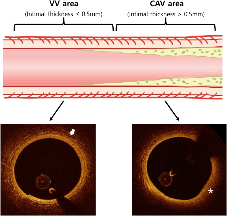
Figure 1.
This illustration shows the study segment of the study. In the VV area, which is adjacent to the CAV area, the VV can be clearly evaluated with OCT (arrow). In the CAV area, the evaluation of the VV in plaque area (asterisk) is difficult because of the limited penetration power of the OCT. CAV, coronary allograft vasculopathy; VV, vasa vasorum.

