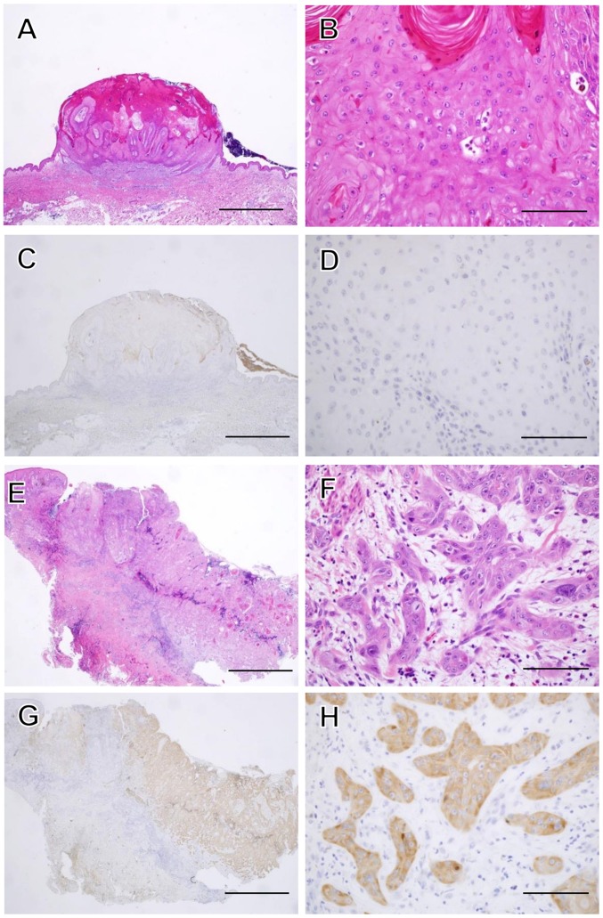
Figure 3.
Histopathological features of KA and SCC. A magnified histological section showing a characteristic architectural pattern (an exo-endophytic lesion with a central keratotic horn) with the involvement of unclear epithelial lips on both sides by the lesions themselves (A). A close-up view of the KA of a lobule made up of large, pale pink cells with a glassy appearance without nuclear atypia (B). IMP3 was not expressed in the KA at all (C and D). A magnified histological section showed an elevated lesion with hyperkeratosis and acanthosis (E). In a close-up view of a SCC section, the neoplastic lobules consisted of squamoid cells, which showed nuclear atypia in the dermis (F). IMP3 was expressed diffusely in the neoplastic lobules of SCC (G and H). Hematoxylin and eosin (H&E) staining was used (A, B, E and F). (C, D, G and H) Sections were stained with anti-IMP3 antibody and peroxidase activity visualized by diaminobenzidine; sections were counterstained with Mayer's hematoxylin. The scale bar is 2 mm (A, C, E and G), and 100 μm (B, D, F and H).

