Figure 5.
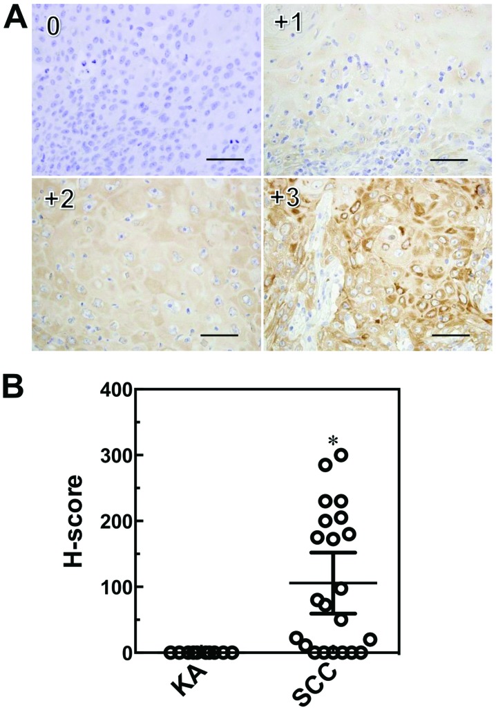
Immunohistochemical scoring system (H-score) for IMP3. Sections were stained with anti-IMP3 and peroxidase activity visualized by diaminobenzidine; sections were counterstained with Mayer's hematoxylin. Staining intensities for IMP3 are shown above (0, +1, +2, +3), with an example each of a membranous and cytoplasmic staining pattern. Scale bars: 50 μm. Immunohistochemical scoring system (H-score) values for IMP3 in SCC and KA tissues. Results are expressed as mean ±95% confidence interval. *P=0.0003, Mann-Whitney U-test compared with KA (B).

