Figure 6.
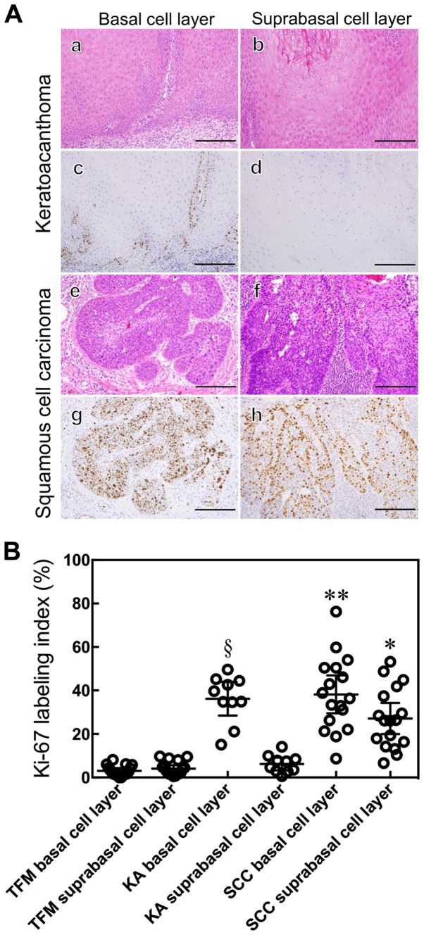
Histological and immunohistochemical staining of KA and SCC. KA (a–d) and SCC (e–h) were examined by either a histological hematoxylin and eosin (H&E) stain (a, b, e and f) or immunohistochemical staining with anti-Ki-67 antibody (c, d, g and h) and peroxidase activity visualized by diaminobenzidine; sections were counterstained with Mayer's hematoxylin. Ki-67 expression was localized in the basal cell layer of the tumor nest in KA (c and d) and in the whole layer of the tumor nest in SCC (g and h). Scale bars: 50 μm (A). The Ki-67 labeling index was determined. Results are expressed as mean ±95% confidence interval. *P<0.0001 compared with the suprabasal cell layer of KA and TFM, Mann-Whitney U-test. **P=0.0445 compared with the suprabasal cell layer of SCC, **P=0.8428 compared with the basal cell layer of KA, Mann-Whitney U-test. §P<0.0001 compared with the suprabasal of KA and basal cell layer of TFM, Mann-Whitney U-test (B).

