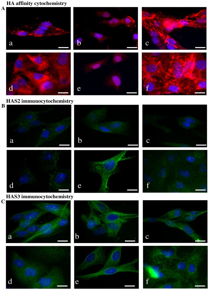
Figure 3.
Detection of HA, HAS enzymes in WM35 and HT168 melanoma cell lines. (A) HA affinity cytochemistry demonstrating secreted HA in red (Streptavidin-Alexa 555). Untreated cells of WM35 (a), WM35 cells treated with 2 μM of CsA (b), and with 5 μM of PD098059 (c), untreated HT168 cells (d), HT168 cells treated with 2 μM of CsA (e), and with 5 μM of PD098059 (f). DAPI was used for visualisation of the nuclei. Scale bar, 20 μm. Representative photomicrographs of 3 independent experiments. (B and C) Immunocytochemistry of HAS2 and HAS3, positive signals appear in green (Alexa 488). Untreated cells of WM35 (a), WM35 cells treated with 2 μM of CsA (b), with 5 μM of PD098059 (c), untreated HT168 cells (d), HT168 cells treated with 2 μM of CsA (e), and with 5 μM of PD098059 (f). DAPI was used for visualisation of the nuclei. Scale bar, 20 μm. Representative photomicrographs of three independent experiments are shown.

