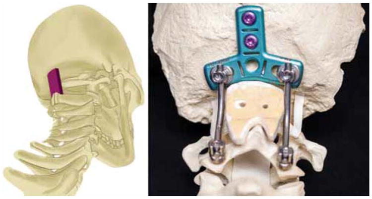
Fig. 1.

Left: 3D illustration demonstrating the interpositional relationship of the graft material between the occiput and C-2. Copyright Department of Neurosurgery, University of Utah. Published with permission Right: Photograph depicting the contouring and fit of the graft material in the construct; note that the notches of the graft fit atop and alongside the spinous process of C-2. Figure is available in color online only.
