Fig. 3.

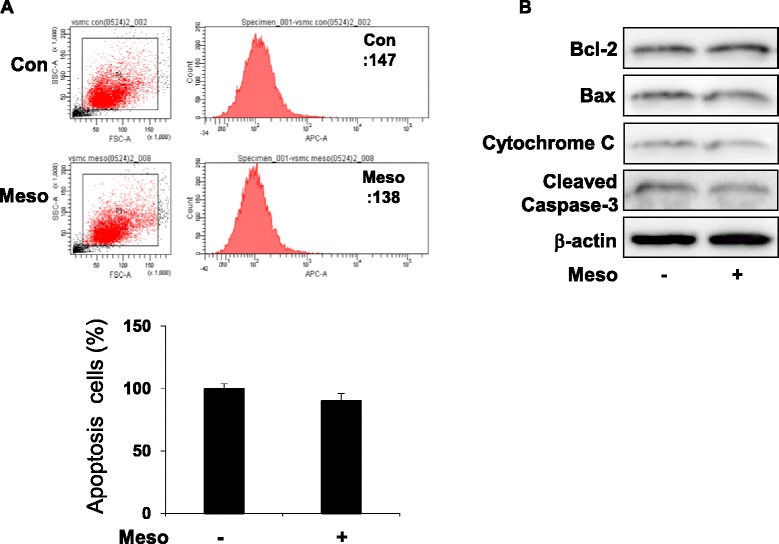
Mesoglycan does not influence apoptotic signaling pathways. Cells were treated with mesoglycan (0.1 μg/ml) for 48 h. Apoptosis was assessed by Annexin V-fluorescein isothiocyanate (FITC) staining by flow cytometric analysis and the percentage of apoptotic cells was then determined (a). Protein expressions of Bcl-2, Bax, cytochrome C and cleaved Caspase-3 were determined by western blot analysis (b). Representative results from three independent experiments are shown
