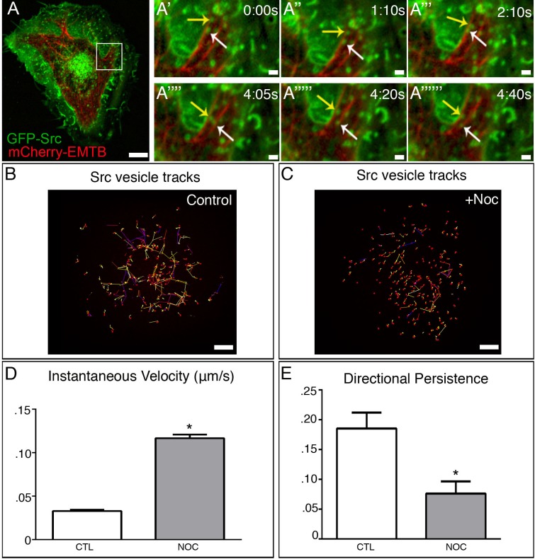
Fig 1. c-Src exhibits bimodal trafficking.
(A) Detection of GFP-Src (green) and 3x-mCherry-EMTB (red) in time-lapse confocal movie of an A7r5 cell. Area in box is enlarged to the right. Bar, 5μm. (A’-A”””) Imaging sequence illustrates that GFP-Src is trafficked bidirectionally along MTs in A. Bar, 2μm (B) GFP-Src vesicles exhibit long, directional movement in time-lapse confocal movie of an A7r5 control (5s/frames). Representative tracks. Bar, 5μm. (C) GFP-Src vesicles exhibit short, randomized movement in time-lapse confocal movie of an A7r5 control (5s/frames). Representative tracks. Bar 5μm. (D) GFP-Src vesicles exhibit increased velocity in response to MT depolymerization. (E) GFP-Src vesicles show reduced directional persistence following nocodazole treatment. N = 5 cells/condition, 50 vesicles/cell. See also S1 Movie. Error bars present SEM. Asterisks indicate p-value < .05.

