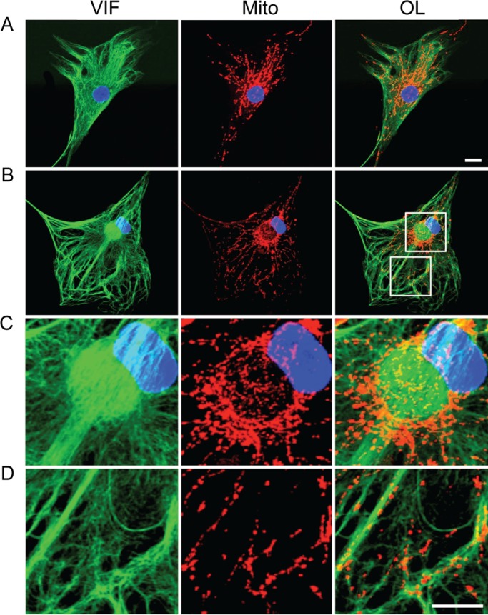
FIGURE 1:

Mitochondria are associated with vimentin IFs. Indirect immunofluorescence with anti-vimentin (VIF), mitochondria stained with MitoTracker Red (Mito), and nucleus stained with Hoechst 33258. (A) Control fibroblasts with typical arrays of vimentin IFs (VIF; green) and containing a normal distribution of mitochondria (Mito; red in the same cell). OL, overlay; blue, nucleus. (B) A GAN fibroblast containing bundles and an ovoid aggregate of vimentin IFs (VIF, green) next to the nucleus (blue); mitochondria in the same cell (Mito, red). Boxed regions in B (OL) are shown at higher magnification to reveal details of mitochondrial association with the aggregate (C) and bundles (D) of vimentin IFs. There are also some mitochondria associated with IFs located between the thicker bundles (D). Confocal images. Scale bars, 10 μm.
