FIGURE 2:
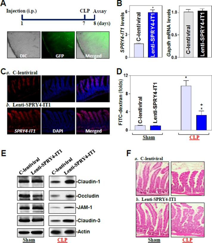
Elevation of mucosal SPRY4-IT1 by infection with a SPRY4-IT1 lentiviral expression vector protects gut TJ barrier function in mice exposed to CLP. (A) Distribution of the lenti-SPRY4-IT1 (GFP) in the small intestinal mucosa 7 d after intraperitoneal (i.p.) injection. (B) Levels of SPRY4-IT1 in the small intestinal mucosa in mice described in A. Values are means ± SEM (n = 4). *p < 0.05 compared with C-control lentiviral vector (C-lentiviral). (C) Distribution of SPRY4-IT1 in the small intestine as measured by FISH in mice described in A. (D) Gut permeability in sham mice and mice exposed to CLP for 24 h. FITC-dextran was given orally, and blood samples were collected 4 h later. *,+p < 0.05 compared with sham or C-lentiviral-treated mice exposed to CLP, respectively. (E) Representative immunoblots of tight junctions in the small intestinal mucosa in mice described in D. (F) Hematoxylin/eosin staining of the intestinal mucosa.

