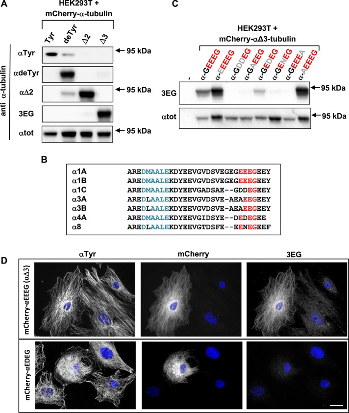
FIGURE 1:
A new antibody specific to the –EEEG protein C-terminus, named 3EG. (A, C) Immunoblot of protein extracts from HEK293T cells expressing different forms of 95-kDa α‑tubulin fused to mCherry at their N-terminus. Expression levels were controlled with αtot antibody. Antibodies recognizing either the unmodified C-terminal α-tubulin tail (tyrosinated tubulin) or its processed versions (detyrosinated or ∆2- or Δ3-tubulin) were used in A, and 3EG antibody was used in C to assay mutation (gray) of the Δ3-tubulin epitope (red). Quantification of the data in A is presented in Supplemental Table S1. (B) Alignment of mouse α-tubulin C-termini. Epitope of αtot antibody in blue; conserved amino acids from the –EEEG C-terminal sequence in red. (D) Immunocytochemistry of MEFs transfected with mCherry–αΔ3-tubulin and a mutated form of this protein ending with EDEG instead of EEEG. Scale bar: 20 µm.

