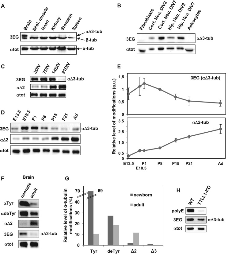
FIGURE 2:
αΔ3-Tubulin, a neuronal variant enriched around mouse birth. Equal quantities of proteins extracted from various tissues and cell types were subjected to immunoblot analysis. Protein levels were controlled using αtot antibody. (A) Immunoblot of crude protein extracts from the indicated neonate mouse tissues. (B) Immunoblot of protein extracts from the indicated cell types, including cortical and hippocampal neurons cultured 2 or 7 DIV. (C) Immunoblot of hippocampal neurons at different stages of culture. (D) Immunoblot of crude protein extracts from mouse brains at different stages of development, including E13.5 and E18.5, postnatal days 1, 8, 15 and 21 (P1–21), and adult (Ad). Mixes of four or five half-brains were used at each developmental stage. (E) Quantitative analysis of three immunoblots for αΔ3 and of five immunoblots for αΔ2 realized as in D with brain protein extracts. In each immunoblot, values obtained were normalized to the value obtained at P8. Error bars indicate SEM; a.u., arbitrary units. (F) Immunoblot of protein extracts from neonate (P1) and adult mouse brains (same extracts as in D). These samples were coanalyzed with extracts from HEK293T cells transfected with the various mCherry–α-tubulin variants (Supplemental Figure S2). (G) Quantitative analysis of immunoblots such as those presented in Supplemental Figure S2. Mixtures of five neonate brains and five adult half-brains were analyzed in two series of Western blots. The plotted values represent the percentages of the different forms of α-tubulin in brains estimated after normalization to total α-tubulin levels (with αtot antibody) and to antibody sensitivity (using modified mCherry–α-tubulins) as explained in Materials and Methods. (H) Immunoblot of crude protein extracts from wild-type (WT) and TTLL1-knockout mouse brains.

