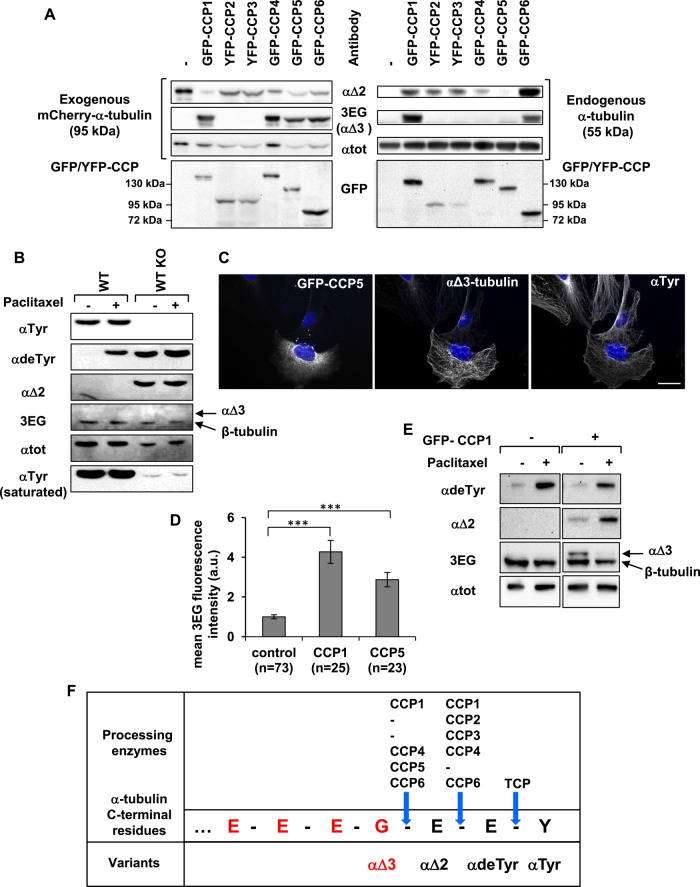
FIGURE 3:
Specificity of CCP enzymes in producing αΔ2 and αΔ3. (A) Immunoblot of protein extracts from HEK293T cells coexpressing each GFP- or yellow fluorescent protein (YFP)–tagged CCP and mCherry–αΔ2-tubulin. Analysis of mCherry–α-tubulin (left) and endogenous α‑tubulin (right). (B) Immunoblot of protein extracted from WT and TTL-knockout (TTL KO) fibroblasts after incubation with dimethyl sulfoxide (DMSO: control) or paclitaxel (15 μM) for 2 h. TTL KO cells contain high levels of detyrosinated and αΔ2-tubulin, but no αΔ3-tubulin is detected. The reactive 3EG band corresponds to β-tubulin (Figure 4). (C) Immunocytochemistry of TTL KO fibroblasts transfected with GFP-CCP5 and immunostained with anti–αTyr-tubulin and 3EG antibody. Scale bar: 20 µm. CCP5 leads to the formation of αΔ3-tubulin. (D) Quantitative analysis of immunocytochemistry experiments as in C using TTL KO fibroblasts transfected with either GFP-CCP1 or GFP-CCP5. Fluorescence was measured as explained in Materials and Methods. (E) Immunoblot of protein extracted from HEK293T cells expressing GFP-CCP1 or not after incubation with DMSO (control) or paclitaxel (50 nM) for 24 h. (F) Schematic representation of the C-terminal amino acids of α1A/B-tubulin, 3EG epitope (red), and processing enzymes associated with the generation of the variants.

