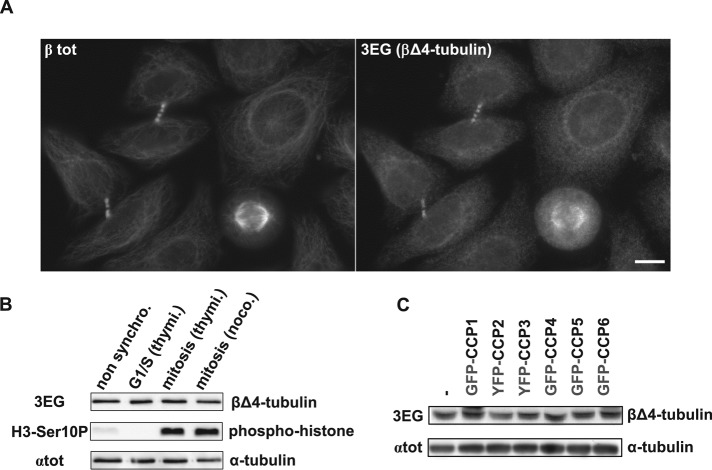
FIGURE 5:
The novel β2A/B-tubulin variant βΔ4 evidenced in HeLa cells by 3EG antibody. (A) Immunocytochemistry of HeLa cells using 3EG and anti–β-tubulin antibody (βtot). Scale bar: 10 µm. (B) Immunoblot of protein extracts from nonsynchronized HeLa cells and from HeLa cells synchronized in G1/S or mitosis (see Materials and Methods). Cell synchronization was verified using a phosphohistone antibody (H3-Ser10P). Loaded proteins were standardized using αtot antibody, and the truncated β-tubulin form was analyzed with 3EG antibody. (C) Analysis of the β-tubulin band in protein extracts from HEK293T cells expressing GFP- or YFP-tagged CCPs (complementary analysis of the experiment in Figure 3A, right, including lower band).

