FIGURE 4:
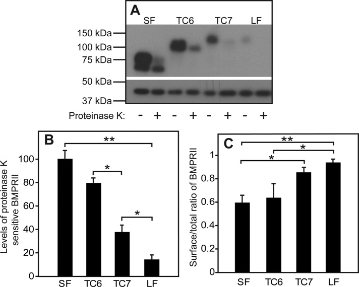
The length of the C-terminal extension of BMPRII-LF determines its steady-state cell surface level. HEK293T cells were transfected as in Figure 1 with the indicated myc-BMPRII constructs. At 24 h posttransfection, the cell surface receptors (exposed to externally added enzyme) were digested (or not) with proteinase K at 4°C (see Materials and Methods). Cells were lysed and subjected to SDS–PAGE and immunoblotting and probing with anti-myc antibodies (A, top) or anti-β-actin (A, bottom; loading control). (A) A representative gel. (B) Quantification of the levels of proteinase K–sensitive myc-BMPRII. Data (mean ± SEM, n = 6) were calibrated relative to the value obtained for myc-BMPRII-SF, taken as 100%. Asterisks indicate significant differences between the bracketed pairs. (C) Ratio of cell surface–localized to total BMPRII levels. The cell surface receptor levels were calculated from the difference between proteinase K–treated and untreated samples, yielding the proteinase K–sensitive fraction. Asterisks indicate significant differences between the bracketed pairs (*p < 0.05; **p < 0.001; Student’s t test).

