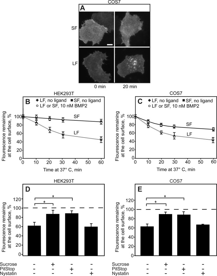
FIGURE 5:
The C-terminal extension of BMPRII-LF directs it to clathrin-mediated endocytosis. HEK293T or COS7 cells were transfected with myc-BMPRII-LF or myc-BMPRII-SF. After 24 h, they were left untreated or subjected to internalization-inhibiting treatments (hypertonic sucrose–supplemented medium, PitStop, or nystatin). The receptors at the cell surface were then labeled at 4°C (time 0) with anti-myc, followed by Alexa 546–GαM Fab’, incubated for defined intervals at 37°C, and fixed at 4°C (Materials and Methods). In experiments conducted in the presence of ligand, BMP2 (10 nM) was added together with the secondary Fab’ and retained throughout the experiment. (A) Typical images. Bar, 10 μm. (B, C) Quantitative endocytosis measurements in HEK293T (B) and COS7 (C) cells. The fluorescence intensity remaining at the cell surface was measured by the point-confocal method (Materials and Methods). Results are mean ± SEM of 200 cells/time point, taking for each sample the intensity at time 0 as 100%. (D, E) Endocytosis of myc-BMPRII-LF is inhibited by CME inhibitors but not by nystatin. For each treatment, the fluorescence intensity at the cell surface was measured at time 0 and after 20 min of incubation at 37°C in medium containing inhibitors (where indicated). Two hundred cells were measured for each condition, and the intensity of the same sample at time 0 was taken as 100%. Treatments that inhibit CME (sucrose and PitStop) induced a significant reduction in myc-BMPRII-LF endocytosis (*p < 0.01).

