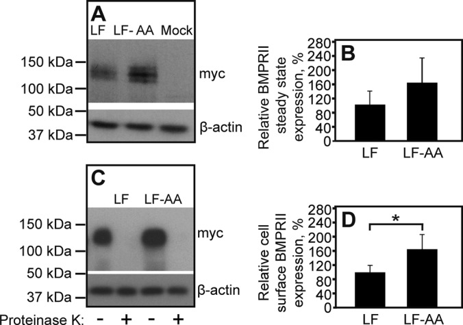
FIGURE 7:

Clathrin-mediated endocytosis attenuates the cell surface expression of BMPRII-LF. HEK293T cells were transfected with myc-BMPRII-LF or myc-BMPRII-LF-AA. At 24 h posttransfection, the steady-state expression of the transfected receptors (A, B) and their expression levels at the plasma membrane (C, D) were measured as in Figures 1 and 4, respectively. (A) A representative gel. (B) Quantification of the steady-state expression levels of myc-BMPRII-LF and myc-BMPRII-LF-AA from multiple experiments (n = 6). Results (mean ± SEM) were normalized relative to β-actin, taking the expression level of myc-BMPRII-LF as 100%. (C) A representative immunoblot with or without proteinase K digestion. (D) Quantification of cell surface–localized myc-BMPRII-LF and myc-BMPRII-LF-AA. Results (mean ± SEM, n = 6) were derived from the difference between proteinase K–treated and untreated samples, as in Figure 4. The asterisk indicates a significant increase (p < 0.05) in cell surface level of myc-BMPRII-LF-AA relative to that of myc-BMPRII-LF (taken as 100%).
