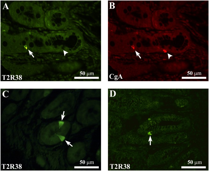
Fig 3. Representative confocal images of T2R38- and CgA-IR cells.
(A) Shows a T2R38-IR cell (arrow) which is immunoreactive for chromogranin A (CgA)-IR (B, arrow) in the colonic mucosa of a NW subject. The arrowheads in A and B indicate a CgA-IR cell not containing T2R38-IR. C and D show the different types of morphology of T2R38-IR cells. Arrows in C point to an “open-type” EEC cell with T2R38-IR, while the arrow in D points to a T2R38-IR cell displaying characteristics of “closed-type” EEC cells. Images in C and D are from colonic mucosa of OW/OB subjects. Both T2R38-IR types of cells were observed also in the colonic mucosa of NW subjects (not shown). Calibration bar: 50μm.

