Fig. 4.

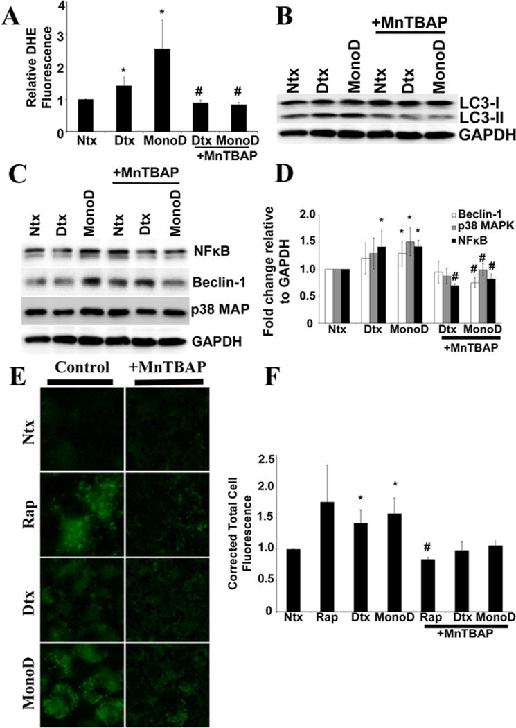
Regulation of autophagic flux and vacuole formation in response to MonoD is mediated by superoxide. A: Cells were pre-treated with 100 μM MnTBAP for 1 h prior to treatment with 50 nM Digitoxin and MonoD. Superoxide production was assayed using DHE fluorescence intensity, respectively, at the peak response time of 15 min post-drug treatment. B: Cells pretreated for 1 h with MnTBAP (100 μM) were treated with 50 nM Digitoxin and MonoD for 1 h and lysed and assayed for detection of autophagy marker LC3-II levels. Blots were re-probed with GAPDH antibody to confirm equal loading of the samples. C: Lysate obtained from cells treated for 1 h with 50 nM Digitoxin and MonoD following 1 h pre-treatment with 100 μM MnTBAP was assayed for autophagy regulatory proteins Beclin-1, NFkB, and p38 MAPK using immunoblotting. D: Beclin-1, NF-κB, and p38 MAPK blots were quantified with densitometry using ImageJ. E: Cells pretreated for 1 h with MnTBAP (100 μM) were treated with 50 nM Digitoxin and MonoD for 1 h and assayed for intracellular autophagic vacuole formation using Cyto-ID®. Following treatment with test drugs, cells were incubated with Cyto-ID® for 30 min at 37°C and intracellular Cyto-ID® fluorescence imaged using EVOS® FL Cell Imaging System at 40× resolution. F: Autophagic vacuoles were quantified using ImageJ after performing background subtraction and plotted as Corrected Total Cell Fluorescence. (*P < 0.05 for each drug treated data point as compared to non-treated control; #P < 0.05 versus Digitoxin- and MonoD-treated datasets).
