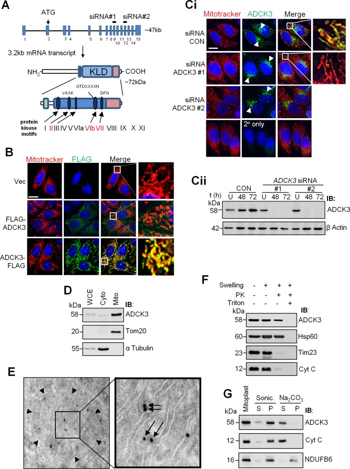
Fig 1. ADCK3 associates with mitochondrial cristae.
(A). Schematic of the ADCK3 gene and its product. The relative position of exons/introns within the ADCK3 gene locus, the initiating ATG codon and the exons targeted by siRNA (ADCK3 siRNA #1 and #2) are depicted. Isoform 1 of ADCK3 is also shown with the relative positions of several important domains and motifs. Green rectangle: Region conserved amongst ADCK family members and specifically related to CoQ biosynthesis. Blue rectangle: Kinase-Like Domain (KLD) which contains 5 of the 12 prototypical kinase motifs involved in ATP binding and the phospohtransfer reaction (Blue ovals). Red rectangle: C-terminal region, whilst highly conserved amongst the ADCK3/4 subgroup, is divergent from that found in classical protein kinases and other ADCK family members. (B). ADCK3-FLAG localises to mitochondria. HeLa cells transiently transfected with pcDNA3.1/Hygro(+) only (Vec) or pcDNA3.1/Hygro(+) containing ADCK3 cDNA with either an N-terminal (pcDNA3.1/Hygro(+)-FLAG-ADCK3: FLAG-ADCK3) or C-terminal (pcDNA3.1/Hygro(+)-ADCK3-FLAG: ADCK3-FLAG) FLAG tag. Immunofluorescence performed with anti-FLAG and Alexa488-conjugated secondary antibodies (FLAG). Counterstaining for mitochondria was performed with Mitotracker Deep Red (Mitotracker). Nuclei were stained with Hoechst 3342. White bars: 15 μm. 63x mag. A single z-axis position is shown. See S1 Fig for images over multiple z-axis positions with ADCK3-EGFP. (C, D). Endogenous ADCK3 localises to mitochondria. Immunofluorescence (C) based analysis of HeLa cells conducted with anti-ADCK3 and Alexa488-conjugated secondary antibodies (ADCK3). HeLa cells treated with control siRNA (siRNA CON) or ADCK3 siRNA #1 and #2 for 48 h. Again, counterstaining for mitochondria was performed with Mitotracker Deep Red (Mitotracker). Nuclei were stained with Hoechst 3342. White bar: 15 μm. Arrow head indicates the position of significant perinuclear or ‘golgi-like’ staining. 63x mag. Cellular subfractionation of HeLa cells (D) was also performed. WCE, cytoplasmic (Cyto) and mitochondrial (Mito) fractions (20 μg) were separated via SDS-PAGE and immunoblotting conducted with antibodies to ADCK3, Tom20 (a mitochondrial marker) and α Tubulin (a cytosolic marker). (E). ADCK3-FLAG localises to mitochondrial cristae. Anti-FLAG immunogold electron microscopy performed with HeLa cells transiently transfected with vector expressing ADCK3-FLAG. Arrow Heads: OMM; Arrows: ADCK3-FLAG on cristae. (F, G). Endogenous ADCK3 associates with the matrix side of the inner mitochondrial membrane. Proteinase K protection (E) and Sonication/Alkaline extraction experiments (F) were conducted with crude mitochondria. Immunoblotting of samples/fractions separated via SDS-PAGE was performed with antibodies to ADCK3, Hsp60 (matrix protein), Tim23 (integral IMM protein facing the IMS), Cyt C (IMM associated IMS protein) and NDUFB6 (integral IMM protein). PK: proteinase K. S: supernatant fraction. P: pellet fraction.

