Fig. 3.

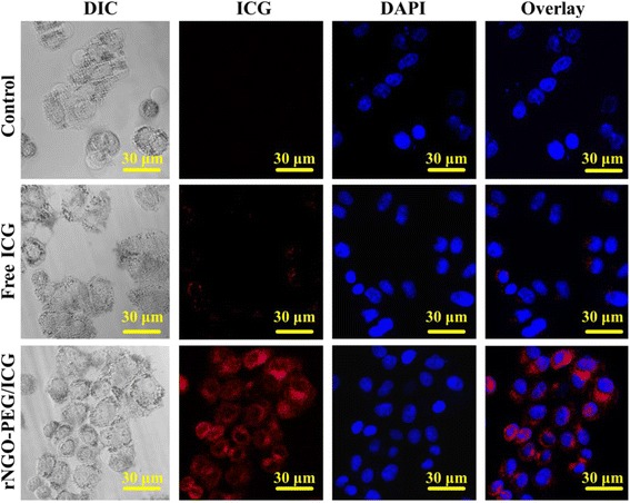
Confocal fluorescence microscopic images of Hela cells treated with PBS (as blank control), free ICG, and rNGO-PEG/ICG for 3 h. The cell nuclei were stained by DAPI

Confocal fluorescence microscopic images of Hela cells treated with PBS (as blank control), free ICG, and rNGO-PEG/ICG for 3 h. The cell nuclei were stained by DAPI