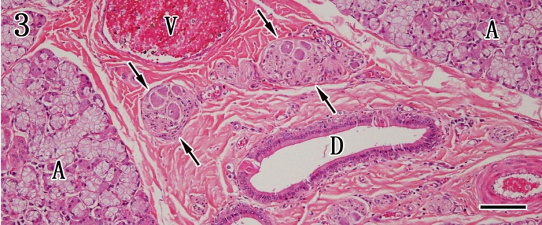
Fig. 3.

Mandibular gland. The mandibular gland is composed of mixed components of mucous and serous acinar cells (A). The positions of the interlobular duct (D), ganglion cells (arrows) and vessels (V) are indicated in the stroma. HE staining. Bar=100 µm.
