
Fig. 7.
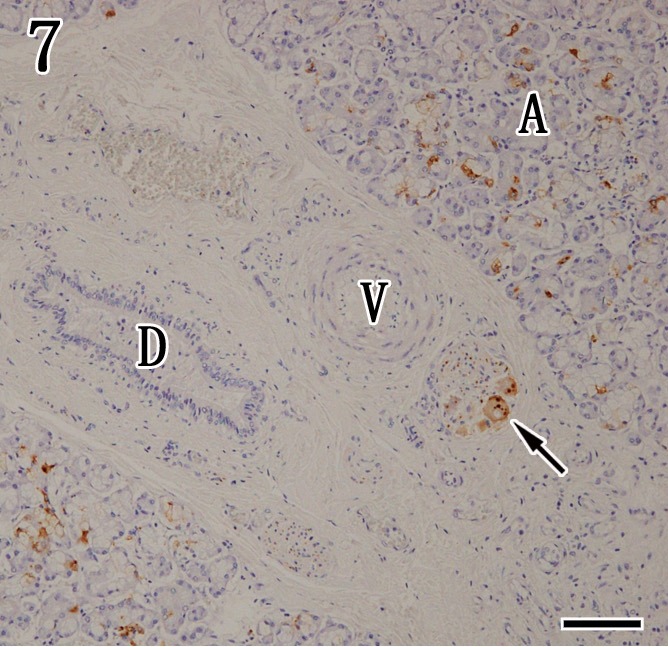
Mandibular gland. Viral antigens were detected by immunohistochemistry with anti-P antibody in the acinar epithelium (A) and ganglion cells (arrow), but the interlobular duct (D) and blood vessels (V) were negative. Immunohistochemistry. Bar=100 µm.