Fig. 10.

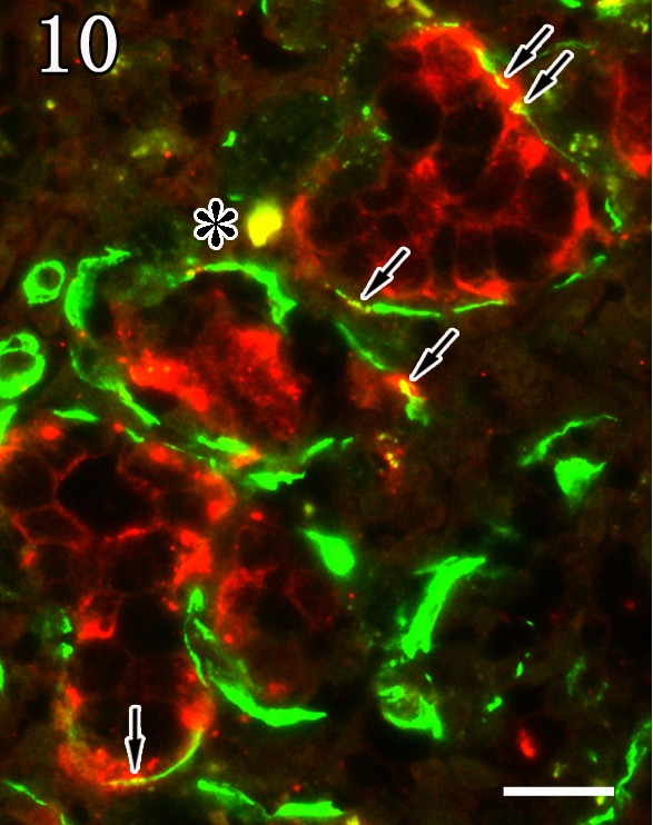
Mandibular gland. Myoepithelial cells were double positive (arrows, yellow) for anti-α-SMA (green) and anti-P antibodies (red). The asterisk indicates an artifact. Double staining. Bar=20 µm.

Mandibular gland. Myoepithelial cells were double positive (arrows, yellow) for anti-α-SMA (green) and anti-P antibodies (red). The asterisk indicates an artifact. Double staining. Bar=20 µm.