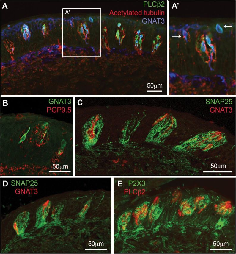
Figure 3.

Double and triple labeling for cell and nerve fiber markers. (A, A′) Horizontal section of the papilla; (B–E) Longitudinal sections where dorsal is to the right. (A, A′) Nerve fibers stained with acetylated tubulin (red) densely innervate taste buds as well as nontaste epithelium between the taste buds. Type II taste cells, marked by staining for PLCβ2 (green) and GNAT3 (Gustducin, blue) populate virtually all taste buds. In addition scattered GNAT3-positive superficial epithelial cells (arrows A′) appear between some taste buds. (B–E) Longitudinal sections of vallate papillae where dorsal is to the right. Note that in this plane of sections, taste buds are oriented at an angle to the surface of the epithelium, so that their taste pore is somewhat more dorsal than the base of the bud. (B) PGP95 stained (red) fibers innervate taste buds as well as surrounding epithelium. Some GNAT3 positive (green) cells appear to contact the PGP9.5-positive nerve fibers. (C–E) The dense network of intragemmal nerve fibers is stained by both SNAP25 (green; C and D) and P2X3 (green; E). These nerve fibers surround and apparently contact the Type II cells marked by GNAT3 (red; C and D) or PLCβ2 (red; E).
