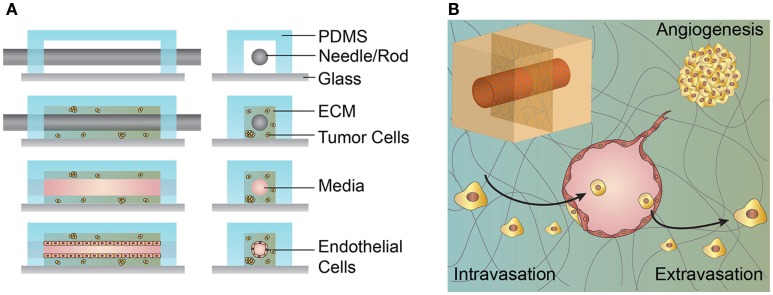
Figure 4.
Schematic illustration of the microvessel fabrication process and interactions between the microvessel and tumor cells in the surrounding extracellular matrix (ECM). (A) A solution form of ECM, often collagen type I or fibrin, laden with cells is introduced around the cylindrical template within the PDMS housing. After gelation/cross-linking, the template rod is removed. Endothelial cells are introduced and line the interior of the cylindrical channel. (B) Upper left inset, cylindrical channel lined with endothelial cells embedded within an ECM. The figure shows a cross-section of a cylindrical vessel interacting with tumor cells in multiple ways. Tumor cells may secrete growth factors and cytokines that promote angiogenesis from a nearby vessel. Tumor cells may invade and intravasate within the local vasculature. Tumor cells within the circulating media may extravasate by adhering to the vessel wall, transmigrating across the endothelium, and invading into the ECM.

