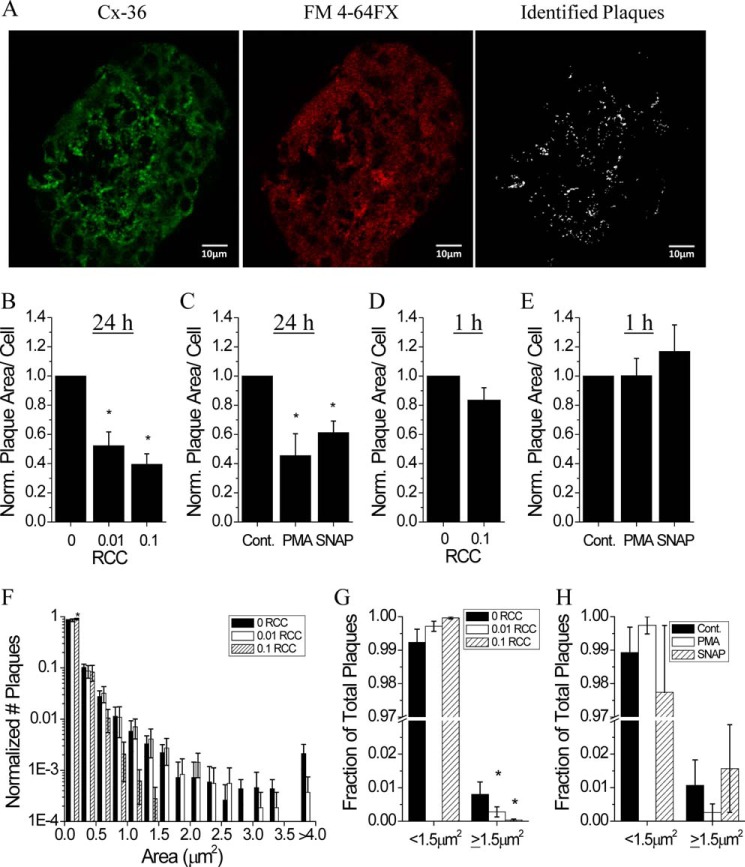
FIGURE 8.
Cytokine-mediated disruption to membrane-localized Cx36 gap junction plaques. A, representative images of a mouse islet immunostained with Cx36, the fixable membrane stain FM 4-64FX, and the identified plaques used for analysis. Cx36 plaque area per cell following treatment with: B, 0, 0.01, or 0.1 RCC for 24 h; C, untreated, 300 nm of the PKCδ activator PMA, or 5 mm of the NO donor SNAP for 24 h; D, 0 or 0.1 RCC for 1 h; E, untreated, PMA, or SNAP for 1 h. Data are normalized to plaque area per cell measured in control islets for each mouse. F, histogram of Cx36 plaque areas for the indicated cytokine treatment. Data are normalized to the total number of plaques measured in control for each mouse. G, fraction of total plaques with an area <1.5 μm2 or ≥1.5 μm2 after treatment with 0, 0.01, or 0.1 RCC for 24 h. H, fraction of total plaques with an area <1.5 μm2 or ≥1.5 μm2 in untreated islets (Cont.) or islets treated with 300 mm PMA or 5 mm SNAP for 24 h. *, indicates a significant difference from control islets based on a 95% confidence interval. Data in B–H are averaged over islets from n = 3–6 mice. Error bars on all panels represent S.E.

