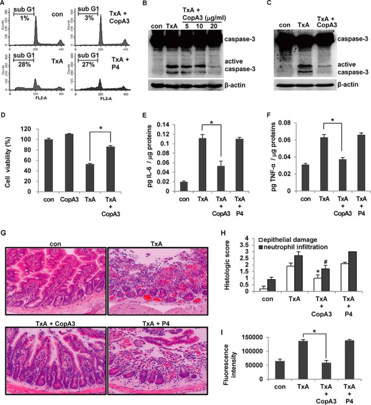
FIGURE 3.
CopA3 inhibits C. difficile toxin A-induced cell toxicity and mouse enteritis. A, HT29 cells (105 cells/well) were pretreated with CopA3 (20 μg/ml) or P4 (20 μg/ml) for 1 h and then incubated with medium (con), toxin A alone (TxA, 3 nm), toxin A plus CopA3, or toxin A plus P4 for 72 h. Cells with fragmented DNA were quantified by PI staining and FACS. The presented results are representative of three independent experiments. B, cells were pretreated with CopA3 for 1 h and incubated with medium (con) or 3 nm toxin A for 72 h. Cell lysates were resolved by 15% SDS-PAGE, and blots were probed with antibodies against caspase-3 and β-actin. C, HT29 cells were pretreated with CopA3 for 1 h, residual CopA3 was removed by washing, and the cells were exposed to toxin A for 72 h. D, cells were pretreated with CopA3 for 1 h and then incubated with toxin A, or toxin A plus CopA3 for 48 h, and cell viability was measured by MTT assay. *, p < 0.001. E–I, male CD1 mice (n = 10/group) were given drinking water containing CopA3 (2 μg/ml) or P4 (2 μg/ml) for 7 days, and then ileal loops were prepared and exposed to 100 μl of PBS (con) or 100 μl of PBS containing toxin A (TxA, 3 nm) for 4 h. The concentrations of IL-6 (E) and TNF-α (F) were measured by ELISA. The bars represent the means ± S.E. from three independent experiments performed in triplicate (*, p < 0.005). G, light micrographs of mouse ileum (hematoxylin and eosin staining, ×200). H, histological scores. *, p < 0.005; #, p < 0.001 compared with mice that received toxin A alone. I, in the mouse enteritis models mentioned above, mucosal macromolecular permeability was measured. The results represent the means ± S.E. from three experiments performed in triplicate (n = 8/group, *, p < 0.005).

