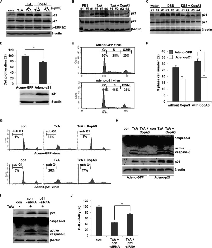
FIGURE 5.
CopA3 reduces p21Cip1/Waf1 expression in colonic epithelial cells. A, HT29 cells were pretreated with CopA3 (10 or 20 μg/ml) or P4 (20 μg/ml) for 1 h and incubated with medium (con), toxin A (TxA, 3 nm) alone, toxin A plus P4, or toxin A plus CopA3 for 72 h. Cell lysates were resolved by 15% SDS-PAGE, and blots were probed with antibodies against p21Cip1/Waf1, p27Kip1, phospho-ERK1/2, and β-actin. B, immunoblot analysis was also performed using proteins extracted from ileal loops of the C. difficile toxin A-induced enteritis model described in Fig. 3. C, immunoblot analysis was performed using proteins extracted from colon samples of the DSS-induced colitis model described in Fig. 4. The presented results are representative of three independent experiments. D, HT29 cells (105 cells/well) were infected with a p21Cip1/Waf1-expressing adenovirus (1 × 10⁷ PFU/ml) or a control GFP adenovirus (1 × 10⁷ PFU/ml) for 24 h, and cell proliferation was assessed using BrdU cell proliferation assays. *, p < 0.001. E, cell cycle distribution was analyzed by PI staining and FACS. F, cells were infected with the above-described viruses for 24 h and then incubated with or without CopA3 (20 μg/ml) for 48 h, and cell proliferation was measured by PI staining and FACS analysis. *, p < 0.01. G, cells were infected with the viruses and then incubated with medium, toxin A alone, or toxin A plus CopA3 (20 μg/ml) for 48 h. Apoptosis was measured by PI staining and FACS analysis. H, cells were infected with the viruses for 24 h and then incubated with medium, toxin A, or toxin A plus CopA3 for 48 h. I, cells were transfected with control siRNA or p21Cip1/Waf1 siRNA for 24 h and then incubated with toxin A for 48 h. J, the viability of the cells described in I was measured by MTT assay. *, p < 0.001.

