FIGURE 6.

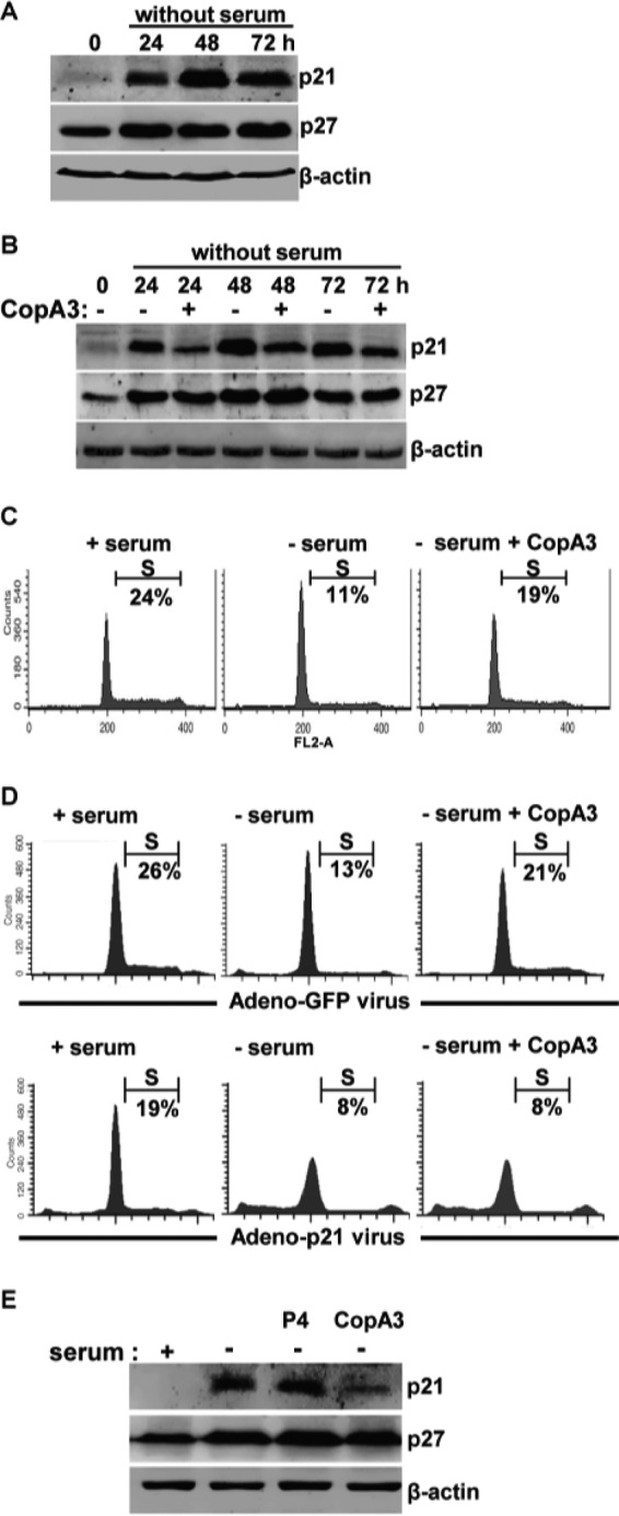
CopA3 restores the growth inhibition caused by serum starvation. A, HT29 cells were incubated in serum-free medium for 24, 48, and 72 h. Cell lysates were resolved by 15% SDS-PAGE, and blots were probed with antibodies against p21Cip1/Waf1, p27Kip1, and β-actin. B, HT29 cells were incubated with medium (0 h), serum-free medium, or serum-free medium plus CopA3 (20 μg/ml) for 24, 48, and 72 h. C, HT29 cells were incubated with medium (+ serum), serum-free medium (− serum), or serum-free medium plus CopA3 (20 μg/ml; − serum + CopA3) for 48 h, and the cell cycle distribution was analyzed by PI staining and FACS. D, HT29 cells were infected with a p21Cip1/Waf1-expressing adenovirus (lower panels) or a control GFP-expressing adenovirus (upper panels) for 24 h and then incubated with medium, serum-free medium, or serum-free medium plus CopA3 for 48 h. The presented results represent the means ± S.E. from three experiments performed in triplicate. E, colonic explants of mice were incubated with medium, serum-free medium, serum-free medium plus P4 (20 μg/ml), or serum-free medium plus CopA3 (20 μg/ml) for 48 h. Cell lysates were resolved by 15% SDS-PAGE, and blots were probed with antibodies against p21Cip1/Waf1, p27Kip1, and β-actin.
