
FIGURE 4.
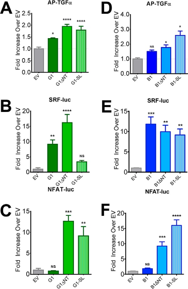
G1-SL exhibits differential levels of signaling activity in distinct assays whereas B1-SL is consistently active. G1-SL exhibited significant signaling activity in the TGFα-shedding (A) and NFAT luciferase (C) assays but was found to not be significantly active in the SRF-luciferase assay (B). However, B1-SL was significantly active at comparable levels to B1ΔNT in all three assays (D: TGFα-shedding, E: SRF-luc, F: NFAT-luc) demonstrating the dispensability of the B1 post-cleavage stalk. All experiments performed in HEK cells. TGFα, SRF-luc, and NFAT-luc results are from 4–6 independent experiments (± S.E. shown, *, p < 0.05; **, p < 0.01; ***, p < 0.001; ****, p < 0.0001 versus cells transfected with empty vector, denoted by EV).