FIGURE 5.
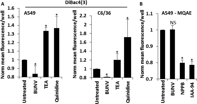
BUNV hyperpolarises the membrane potential through K+ channel activation. A (i) A549 or (ii) C6/36 cells infected with BUNV (MOI 1), or treated with TEA (10 mm)/quinidine (200 μm) were exposed to the membrane potential sensitive fluorescent dye DiBAC4 (3) (20 μm). Values represent 6 hpi/treatment and are normalized to untreated controls (n = 3). Results are expressed as the mean fluorescence per well ± S.E. of averaged widefield images (*, significant difference from untreated cells at the p ≤ 0.05 level). Data are representative of at least three independent experiments. B, A549 cells infected with BUNV (MOI 1) or treated with NPPB (10 μm)/IAA-94 (100 μm) were exposed to MQAE. Fluorescence was acquired as in Fig. 5A. Values represent 6 hpi/treatment and are normalized to untreated controls (n = 3). Results are expressed as the mean fluorescence per well ± S.E. of averaged widefield images (*, significant difference from untreated cells at the p ≤ 0.05 level). Data are representative of three independent experiments.

