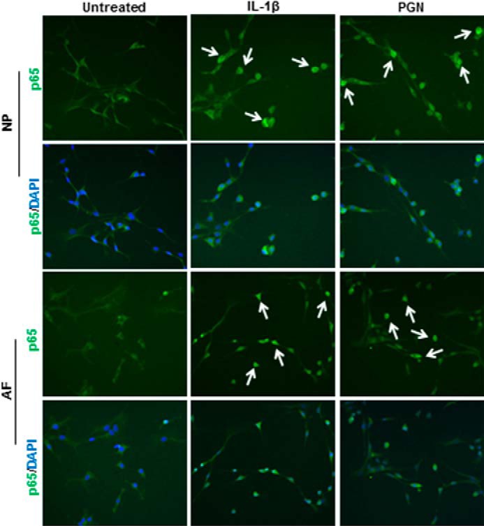
FIGURE 5.

p65 Translocation to the cell nucleus. Cells were treated with IL-1β, PGN, or left untreated or left untreated for 1 h and then stained for p65 localization (green). Cultures were counterstained with the nuclear DNA stain DAPI (blue). White arrows indicate examples of p65 translocation to the nucleus. n = 3.
