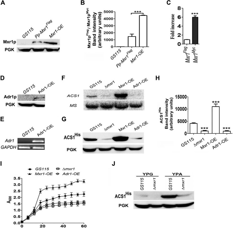
FIGURE 2.
The effect of overexpression of Mxr1p and Adr1p on ACS1 expression and growth of cells in YPA medium. A, comparison of Mxr1p levels in GS115, Pp-Mxr1FLAG expressing Mxr1p from its own promoter and Mxr1-OE overexpressing Mxr1p from the GAPDH promoter. Mxr1p was detected using anti-FLAG/anti-c-myc antibodies. Phosphoglycerate kinase (PGK) levels served as a loading control. B, quantification of the data in A. The intensity of individual bands was quantified and expressed as arbitrary units ± S.D. relative to controls. Data are the average of three independent experiments. ***p < 0.0005. C, analysis of Mxr1 mRNA levels by qPCR. Error bars indicate mean ± S.D. ***, p < 0.0005. One-way analysis of variance, followed by Tukey's multiple comparison test was done (n = 3). D, Western blotting analysis of Adr1p in the Adr1-OE strain using anti-c-myc antibodies. PGK levels served as a loading control. E, RT-PCR analysis of Adr1 mRNA in the GS115 and Mxr1-OE strains. F, Northern blotting analysis of ACS1 expression. MS encoding methionine synthase was used as a loading control. G, analysis of ACS1His levels by Western blotting using anti-His antibodies in different P. pastoris strains. PGK served as a loading control. H, quantification of the data in G. The intensity of individual bands was quantified and expressed as arbitrary units ± S.D. relative to controls. Data are the average of three independent experiments. ***, p < 0.0005. I, growth curves of different P. pastoris strains cultured in YPA medium. Error bars indicate mean ± S.D. (n = 3). J, analysis of ACS1His levels by Western blotting using anti-His antibodies in cells cultured in YPG and YPA containing glycerol and acetate as carbon sources, respectively. PGK served as a loading control.

