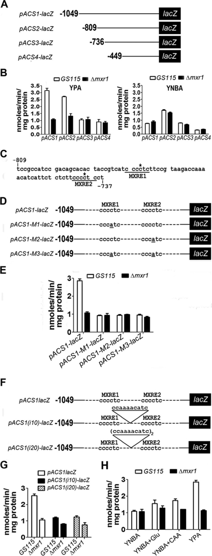
FIGURE 4.

Study of lacZ expression from pACS and identification of pACS-MXREs. A, schematic of pACS-lacZ constructs. B, estimation of β-galactosidase activity in lysates of cells transformed with pACS-lacZ constructs. Cells were cultured in YPA or YNBA. C, nucleotide sequence of pACS between −809 and −737 bp. MXRE1 and MXRE2 are underlined. The cytosine residue within the MXRE crucial for Mxr1p binding is indicated by an asterisk. D, schematic of pACS1-lacZ constructs containing wild-type and mutant MXREs. Point mutations within MXREs are underlined. E, estimation of β-galactosidase activity in lysates of cells transformed with pACS-lacZ constructs containing wild-type and mutant MXREs. F, schematic of pACS1-lacZ constructs carrying 10- or 20-bp insertions between the two MXREs. G, effect of insertion of 10- and 20-bp insertions between the two MXREs on β-galactosidase activity in lysates of cells transformed with pACS-lacZ constructs. β-Galactosidase activity measurements represent the mean ± S.D. of data from three independent experiments. H, lacZ expression from pACS1 in cells cultured in various media as indicated.
