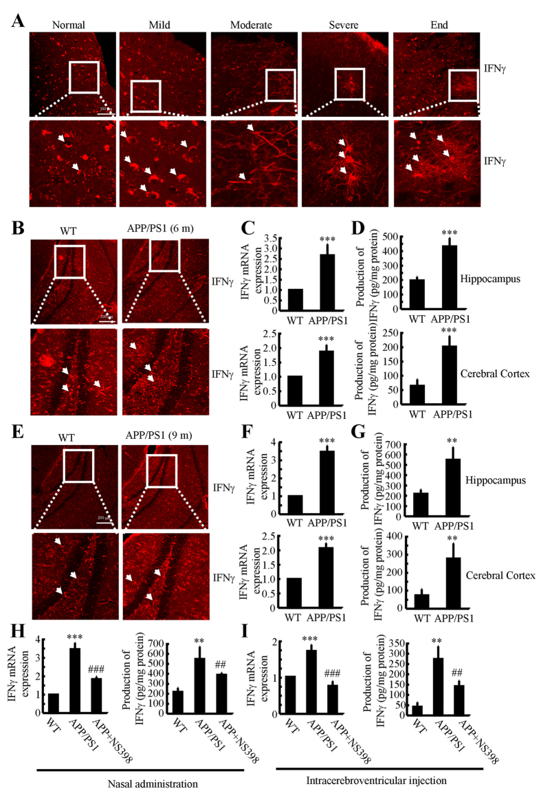
Figure 1. NS398 treatment decreases the induction of IFNγ in APP/PS1 mice.
(A) The tissue blocks of human brains at different stages of AD were collected by the New York Brain Bank at Columbia University. Free-floating slices (40 μm) were prepared by cryostat. (B–G) The brains of WT or APP/PS1 transgenic mice at 6 or 9 months of age were collected following anesthesia and perfusion. In select experiments, the APP/PS1 transgenic mice at the age of 3 month received NS398 (50 μg/kg/d) intranasally for 6 months before brain harvesting (H). In separate experiments, APP/PS1 mice were injected (i.c.v) with NS398 (2 μg/5 μl) for 24 h (I). The immunoreactivity of IFNγ was determined by immunohistochemistry using an anti-IFNγ antibody (A,B,E). The arrows demonstrated the positive staining of IFNγ. IFNγ protein and mRNA levels were determined by IFNγ enzyme immunoassay kits and qRT-PCR, respectively (C–I). Total amounts of protein and GAPDH served as an internal control. The data represent the means ± S.E. of atleast three independent experiments. *p < 0.05; **p < 0.01 and ***p < 0.001 with respect to WT control. #p < 0.05; ##p < 0.01 and ###p < 0.001 compared to APP/PS1 alone.

